| Difficult | Execution Time | Data Analysis | Radioactive Sources |
|---|---|---|---|
| No | Yes |
Hardware setup
This experiment guide is referred to the SP5700 educational kit.
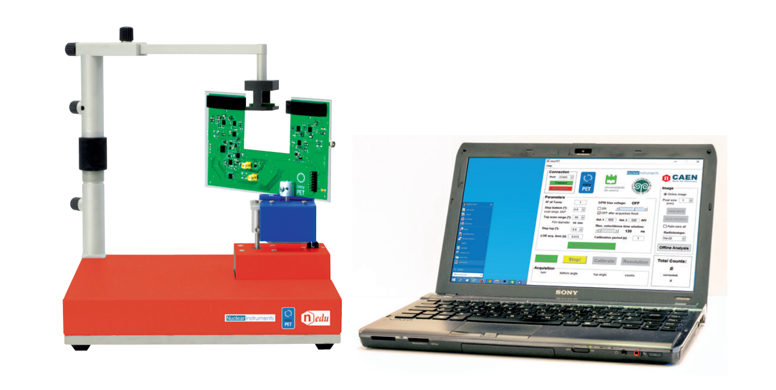
Equipment: SP5700 – EasyPET
| Model | SP5700 | Additional Tool |
|---|---|---|
| Description | EasyPET | 22Na Radioactive source (recommended: 1/2 inch disc, 10 μCi) |
Purpose of the experiment
Understanding the technique of the nuclear imaging and the setup optimization of the parameters by performing two-dimensional image reconstruction of 22Na radioactive source.
Fundamentals
The EasyPET operation principle is simple: two detector modules move together and execute two types of independent movements, around two rotation axes, so as to cover a field of view similar to that of a complete ring of detectors. The rotation movements are executed by two stepper motors. The bottom motor has a fixed axis, whose position defines the center of the field of view. The bottom motor supports and performs a complete rotation of a second motor, in predefined steps of amplitude α. The axis of the top motor is thus always positioned within a circumference of radius equal to the distance between the two axes. The top motor, in its turn, supports and moves a U-shaped printed circuit board, where a pair of aligned and collinear detector modules is mounted, performing a symmetric scan of range θ around the center, for each position of the bottom motor. In this way, EasyPET can reconstruct an image of a radioactive source placed anywhere within a cylindrical field of view between the pair of detectors. The diameter of the field of view is defined by the amplitude of θ, the range of the top motor scan.
Carrying out the experiment
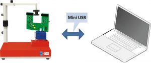
Experimental setup block diagram
- Mount the arm of the source holder on the column fixed on the system base, fix the U–shaped board to the top stepper motor and connect the flat cable to the U-shaped board and to control unit.
- Connect to PC and power ON the system. The parameters involved in the setup optimization for the two-dimensional reconstruction of the source image are three: the detectors operating voltage, the coincidence time window and the threshold.
- Place the source holder between the two detector modules and tune the parameters to estimate the DCR contribution.
- Place the 22Na radioactive source in the holder and repeat the measurement tuning the parameters in order to obtain a good image reconstruction of the radioactive source.
Results
By tuning the parameters, the students can directly observe and understand their effects on the imaging measurements. At fixed threshold, the image contrast changes due to the time window width. The use of the lowest possible coincidence time window of the system is mandatory to achieve a good image contrast. Fixing the bias voltage and the time window, it is interesting to observe how the threshold affects the image contrast. This parameter choice is dictated by a trade off between the signal to noise ratio and efficiency maximization. The threshold value and the coincidence time window should be set by choosing the best compromise.
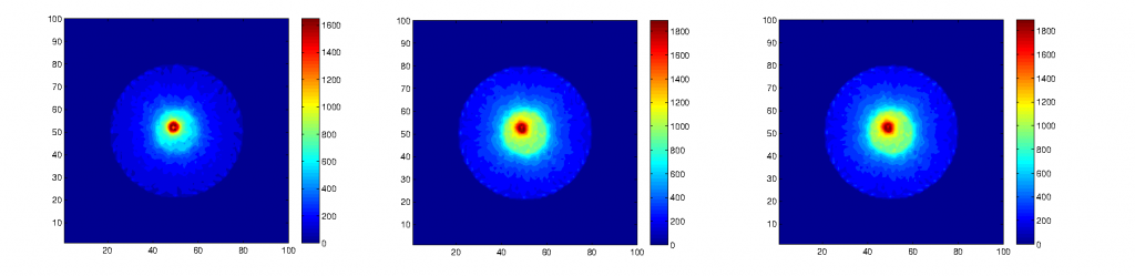
22Na source distribution image as a function of coincidence time window, at fixed threshold and bias voltage