Nuclear Imaging – PET Experiments
Positron Emission Tomography (PET) is one of the most advanced medical imaging techniques, providing detailed information about physiological processes inside the human body. Its development represents a successful integration of different fields, including medicine, nuclear physics, radiochemistry, detector technology, and information processing.
The experiments presented in this section introduce students to the fundamental principles behind PET imaging through a hands-on approach. Using a dedicated educational PET system, students can explore the concepts of positron emission, gamma-ray detection, coincidence measurements, and image reconstruction, gaining insight into the physical processes underlying modern medical imaging techniques.
The proposed activities are performed using the CAEN EasyPET portable educational PET system, developed in collaboration with the University of Aveiro (Portugal). Depending on the experiment, additional data acquisition and analysis instrumentation can be used together with the EasyPET system to investigate the different stages involved in PET measurements.
This experimental approach allows students to understand the basic principles of PET technology and appreciate the role of physics and instrumentation in advanced diagnostic applications.

 |
|
|
|---|---|---|
| SP5701 | SP5700 | |
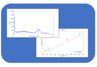
| SG6131 – Basic Measurements: Gamma Spectroscopy and System Linearity | ✔ | |
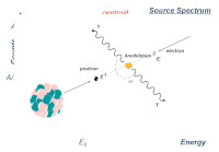
| SG6132 – Positron Annihilation Detection | ✔ | ✔ |
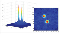
| SG6113 – Two-dimensional Reconstruction of a Radioactive Source | ✔ | ✔ |
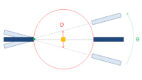
| SG6134 – Spatial Resolution | ✔ | ✔ |
Note: The star indicates the recommended Kits while the yellow color highlights the Kits especially dedicated to the experiences of this Section.